Female Upper Thigh Anatomy - Vastus Medialis Strengthening For Superior Knee Health ... - For more details go to edit properties.
byAdmin•
0
Female Upper Thigh Anatomy - Vastus Medialis Strengthening For Superior Knee Health ... - For more details go to edit properties.. Linea aspera and popliteal surface minimus: Foundational anatomy provides medical students with the necessary background in anatomy for success in clerkships. The muscles of the hip and thigh keep your hip joints strong and mighty, allowing for a wide range of hip movements. Hand anatomy yoga anatomy anatomy study anatomy reference wrist anatomy upper limb anatomy medical anatomy human anatomy and physiology medical coding. To practice tricky questions and answers on all areas of human anatomy, here is complete set of 1000+ multiple choice questions and answers.
For more details go to edit properties. Linea aspera and popliteal surface minimus: Female anatomy includes the external genitals, or the vulva, and the internal reproductive organs. Gluteal tuberosity and upper 1/4 of linea aspera. Free samples of various female poses.
Muscles of the Thigh and Gluteal Region - Part 2 - Anatomy ... from i.ytimg.com Our objective was to describe the muscular and neurovascular anatomy of the medial thigh compartment. Muscles of the anterior thigh. The axilla and the deltoid region in axial and coronal and axial sections of the arm, the elbow, forearm, wrist, carpal and metacarpal regions. Thus, the right side of the image is the patient's left. Gluteal tuberosity and upper 1/4 of linea aspera. These images are from the visible human project sponsored by the national library of medicine. Lymphovenous anastomosis (lva) requires a precise knowledge of the anatomy of the superficial lymphatic collectors in relation to the superficial methods: The final chapter presents anatomical charts of anatomical sections of the upper limb:
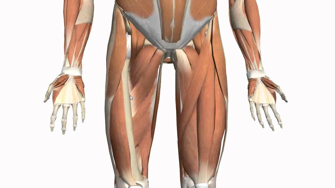
Appendicular muscles of the pelvic girdle and lower limbs.
Foundational anatomy provides medical students with the necessary background in anatomy for success in clerkships. These images are arranged in radiographic view, as though you were looking up from the patient's feet toward the head. The femur is the anatomical name for the thigh bone. Dissections were performed in unembalmed female cadavers. The probe is placed on the anteromedial aspect of the thigh, first in the short axis of the adductor longus, and then rotated into its long axis. Relationships of medial thigh structures were evaluated relative to the midpubic arch and obturator nerve. The female body experiences a greater curvature of the femur to make obvious exception to the female body's wider pelvic region. In human anatomy, the thigh is the area between the hip (pelvis) and the knee. This can effectively educate everyone on the female human body. Mri of upper leg (femur). Gluteal tuberosity and upper 1/4 of linea aspera. Appendicular muscles of the pelvic girdle and lower limbs. There may be variations in treatment that.
This article looks at female body parts and their functions, and it provides an interactive diagram. To practice tricky questions and answers on all areas of human anatomy, here is complete set of 1000+ multiple choice questions and answers. Free samples of various female poses. Anatomically, it is part of the lower limb. 3d human upper leg anatomy or anatomical and muscle set or collection.
Drawn man leg muscle - Pencil and in color drawn man leg ... from art4clip.com This webpage presents the anatomical structures found on thigh mri. This can effectively educate everyone on the female human body. Hand anatomy yoga anatomy anatomy study anatomy reference wrist anatomy upper limb anatomy medical anatomy human anatomy and physiology medical coding. The femur is the anatomical name for the thigh bone. Muscles of the leg and foot. Pictures of upper thigh muscles. Dissections were performed in unembalmed female cadavers. Now that you watched the video, you.
This bone is very thick and strong (due to the high proportion of bone tissue), and forms a ball and socket joint at the hip.
Pictures of upper thigh muscles. Collection by renaud galand • last updated 12 weeks ago. Want to learn more about it? Now that you watched the video, you. Female anatomy includes the external genitals, or the vulva, and the internal reproductive organs. See more ideas about female bodies, anatomy, female anatomy. Anatomy atlases, the anatomy atlases logo, and a digital library of anatomy information are all trademarks of michael p. This can effectively educate everyone on the female human body. Muscles of the leg and foot. Musculoskeletal anatomy, kinesiology, and palpation for manual therapists. Foundational anatomy provides medical students with the necessary background in anatomy for success in clerkships. For more details go to edit properties. Upper thigh anatomy (page 1).
This article looks at female body parts and their functions, and it provides an interactive diagram. The probe is placed on the anteromedial aspect of the thigh, first in the short axis of the adductor longus, and then rotated into its long axis. This can effectively educate everyone on the female human body. Collection by renaud galand • last updated 12 weeks ago. Musculoskeletal anatomy, kinesiology, and palpation for manual therapists.
Posterior Thigh from www.wesnorman.com These images are arranged in radiographic view, as though you were looking up from the patient's feet toward the head. Pelvic & upper thigh anatomy. Want to learn more about it? The single bone in the thigh is called the femur. Our objective was to describe the muscular and neurovascular anatomy of the medial thigh compartment. Dissections were performed in unembalmed female cadavers. Appendicular muscles of the pelvic girdle and lower limbs. Muscles of the leg and foot.
Pictures of upper thigh muscles.
Relationships of medial thigh structures were evaluated relative to the midpubic arch and obturator nerve. Lymphovenous anastomosis (lva) requires a precise knowledge of the anatomy of the superficial lymphatic collectors in relation to the superficial methods: 3d human upper leg anatomy or anatomical and muscle set or collection. Want to learn more about it? Our objective was to describe the muscular and neurovascular anatomy of the medial thigh compartment. Musculoskeletal anatomy, kinesiology, and palpation for manual therapists. Appendicular muscles of the pelvic girdle and lower limbs. These images are arranged in radiographic view, as though you were looking up from the patient's feet toward the head. In human anatomy, the thigh is the area between the hip (pelvis) and the knee. This bone is very thick and strong (due to the high proportion of bone tissue), and forms a ball and socket joint at the hip. The single bone in the thigh is called the femur. The femur is the anatomical name for the thigh bone. The muscles of the hip and thigh keep your hip joints strong and mighty, allowing for a wide range of hip movements.
3d human upper leg anatomy or anatomical and muscle set or collection upper thigh anatomy. The final chapter presents anatomical charts of anatomical sections of the upper limb: